Have you ever wondered how things look from a microscope? Well, you don’t have to anymore. The 2016 Nikon Small World Photography winners were announced. Not only are all the pictures marvelous, but also really impressive.
The photographers can enter to the contest from all over the world, and are given the task to show how beauty can be found in the smallest parts of our world. The photographs must be taken with microphotography, showing impressive technical skills.
Below you will find are the 20 winning pictures chosen by the judges.
1st Place.
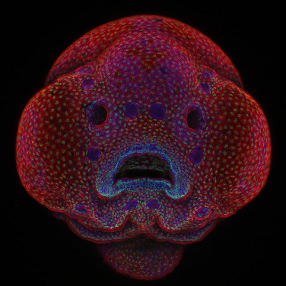
The award goes to Dr. Oscar Ruiz for his picture of four-day-old zebrafish embryo.
2nd Place.
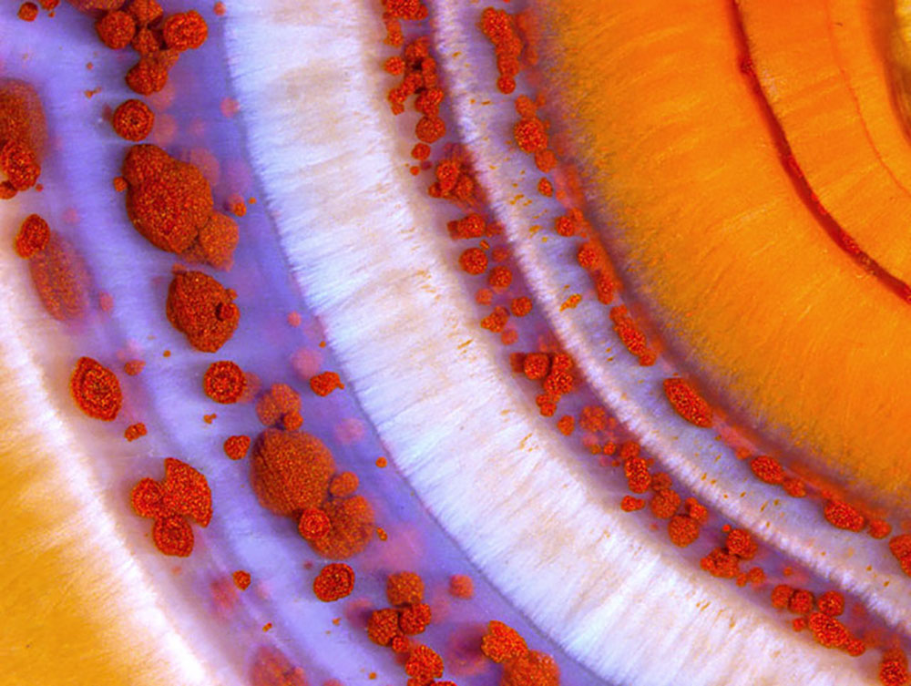
The second place goes to Douglas L. Moore with his piece of polished slab of Teepee Canyon agate.
3rd Place.
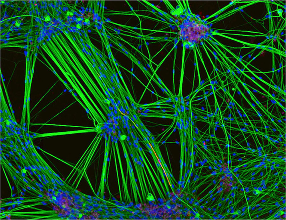
This one goes to Rebecca Nutbrown. Her subject was Brain cells from skin cells: Specifically, this is a culture of neurons (stained green) derived from human skin cells, and Schwann cells, a second type of brain cell (stained red), which have started to cover the neuron in the same way these cells interact in the brain.
4th Place.
This piece was taken by Jochen Schroeder with the Image Stacking technique.
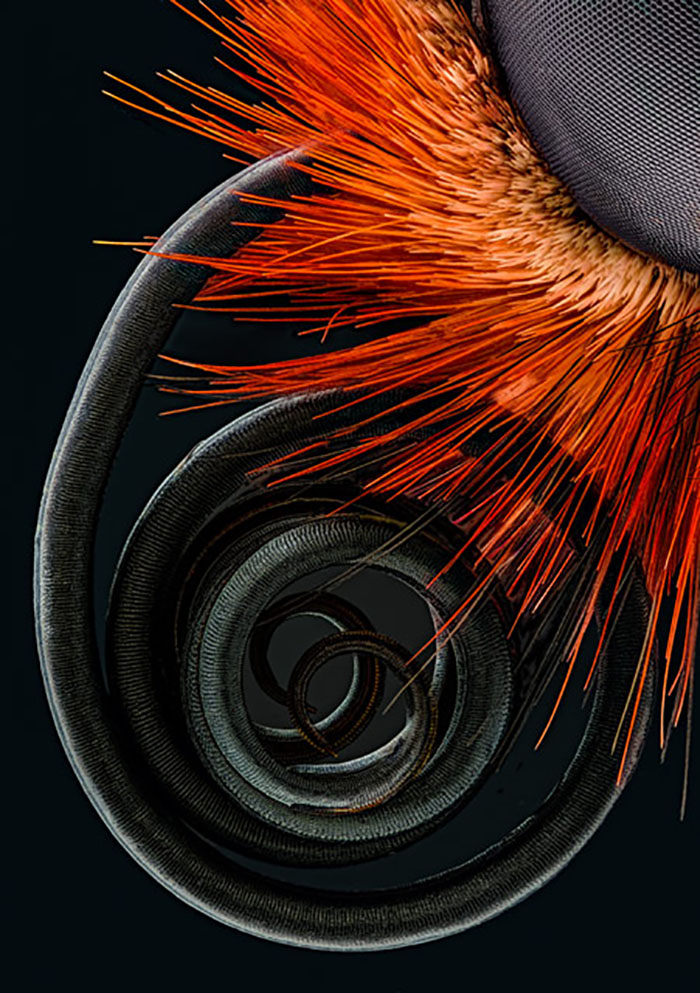
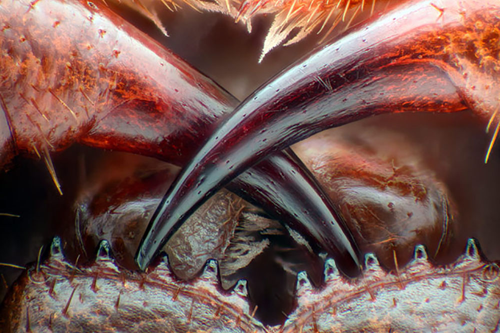
5th Place.
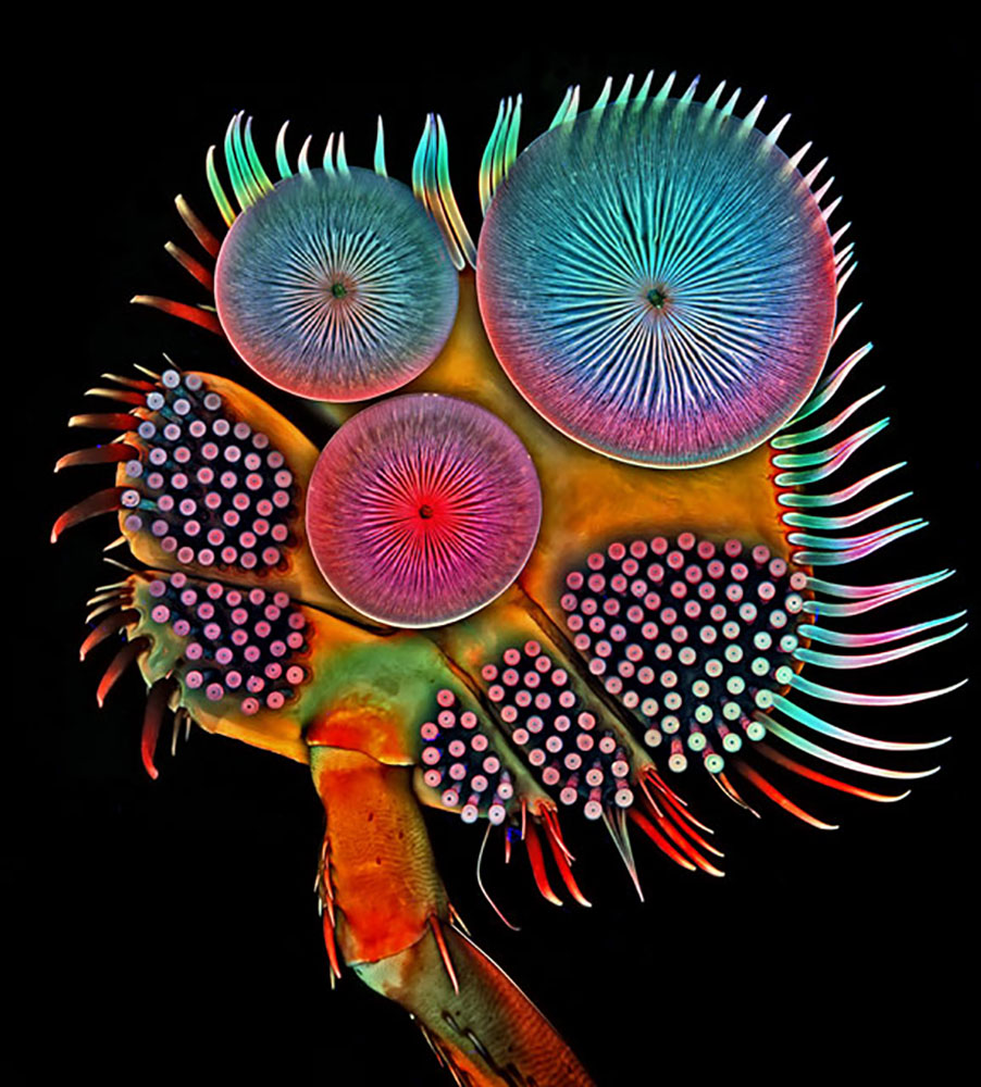
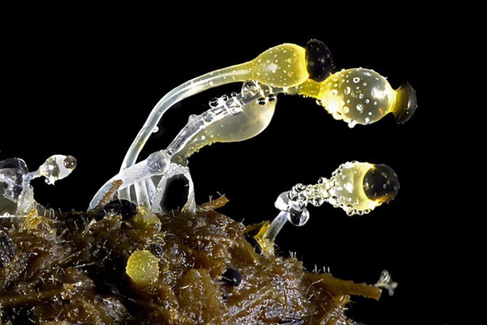
The fifth place goes to Front foot (tarsus) of a male diving beetle taken by Dr. Igor Siwanowicz.
6th Place.

This place is for Marek Mis from Poland.
7th Place.
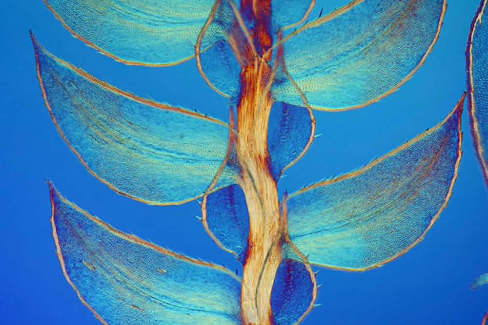
This one belongs to the UK and it’s called Leaves of Selaginella (lesser club moss).
8th Place.
Using the technique of Fiber Optic Illuminatiom this place goes to Samuel Silberman.
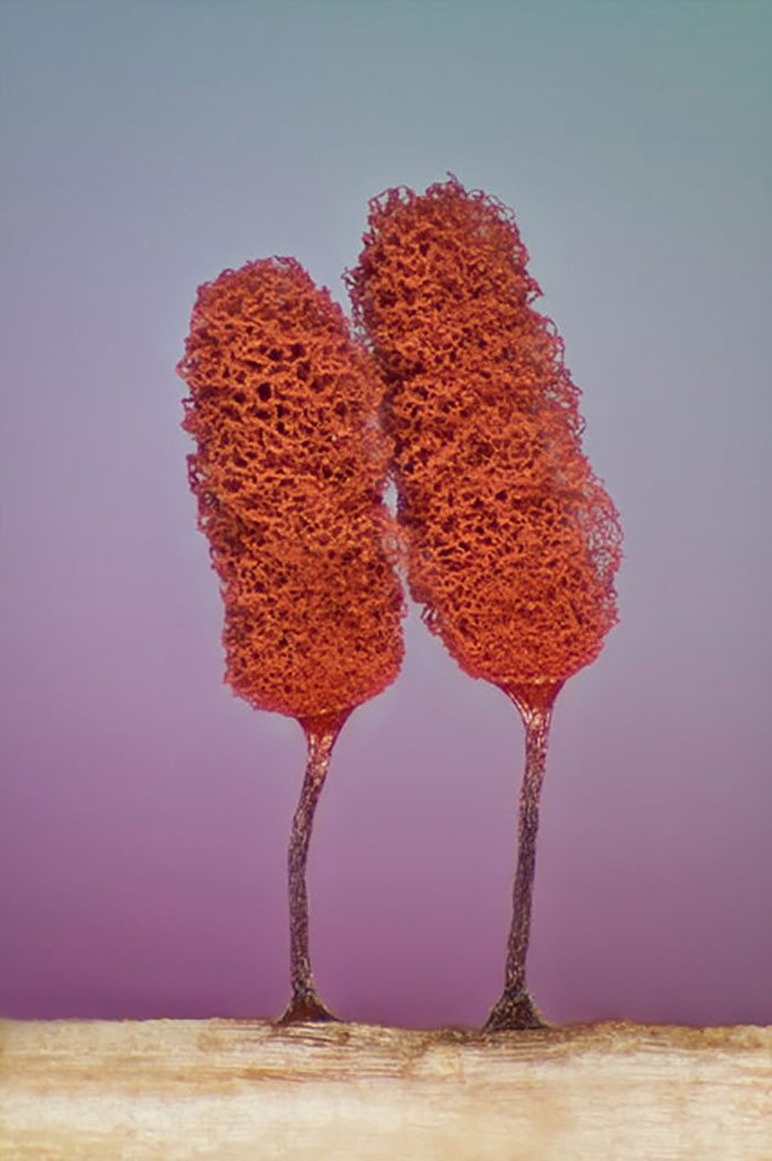
9th Place.
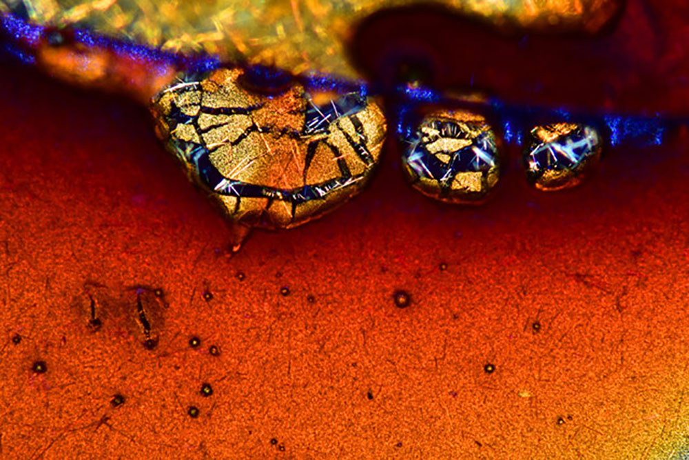
From Japan, Espresso coffee crystals.
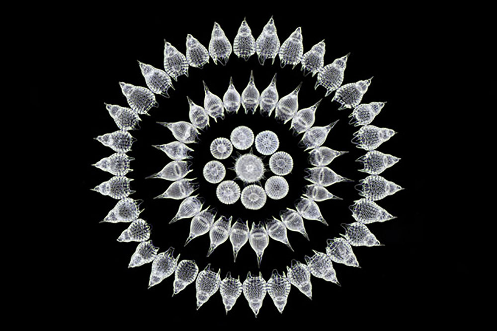
10th Place.
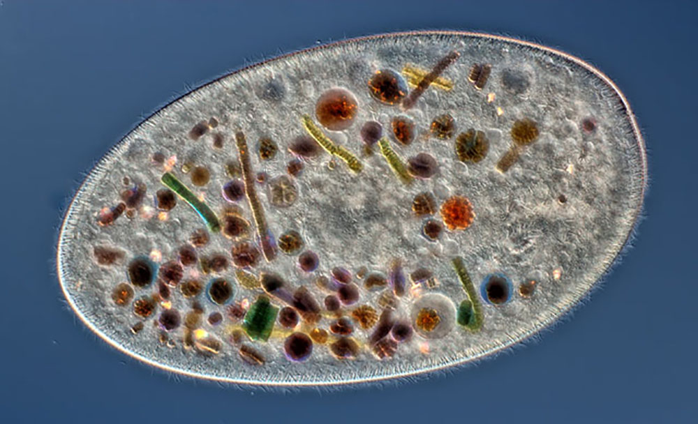
Using the technique of Differential Interference Contrast, Rogelio Moreno Gill from Panama.
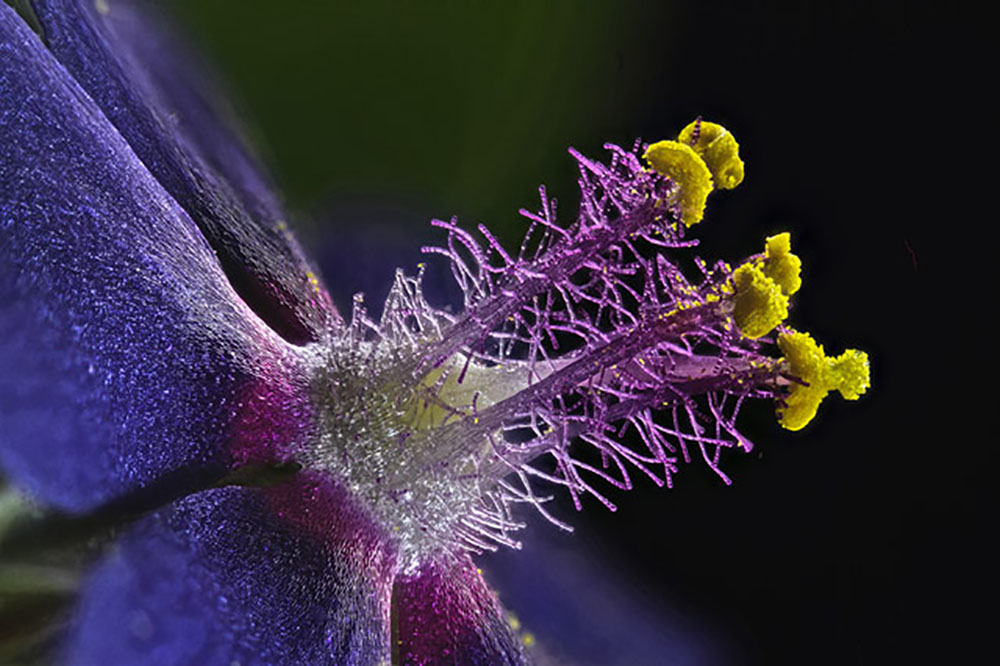
11th Place.
From Belgium using the technique of Macroscopy.
12th Place.
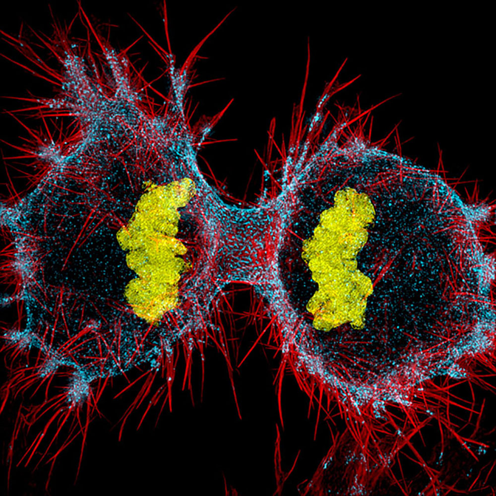
Dr. Dylan Burnette from Vanderbilt University, School of Medicine, Nashville, Tennessee, USA.
13th Place.
This piece was created in Illinois by Walter Piorkowski
14th Place.
Dr. Keunyong Kim took Mouse retinal ganglion cells.
15th Place.
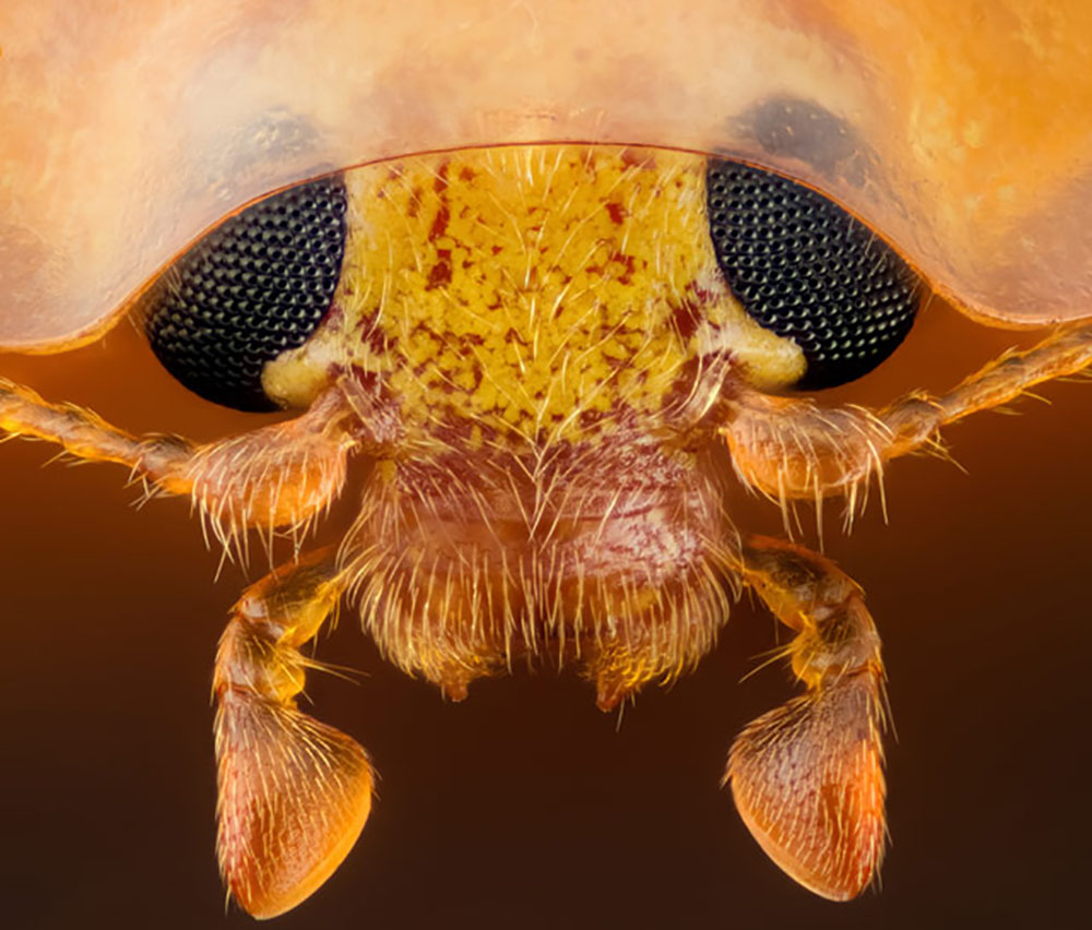
The subject was Head section of an orange ladybird (Halyzia sedecimguttata) with the technique:
Reflected Light, Focus Stacking.
16th Place.
Taken in Itali, Stefano Barone created this with Darkfield.
17th Place.
This was taken at the University of Puerto Rico (UPR), Mayaguez Campus,Biology Department, Mayaguez, Puerto Rico, USA.
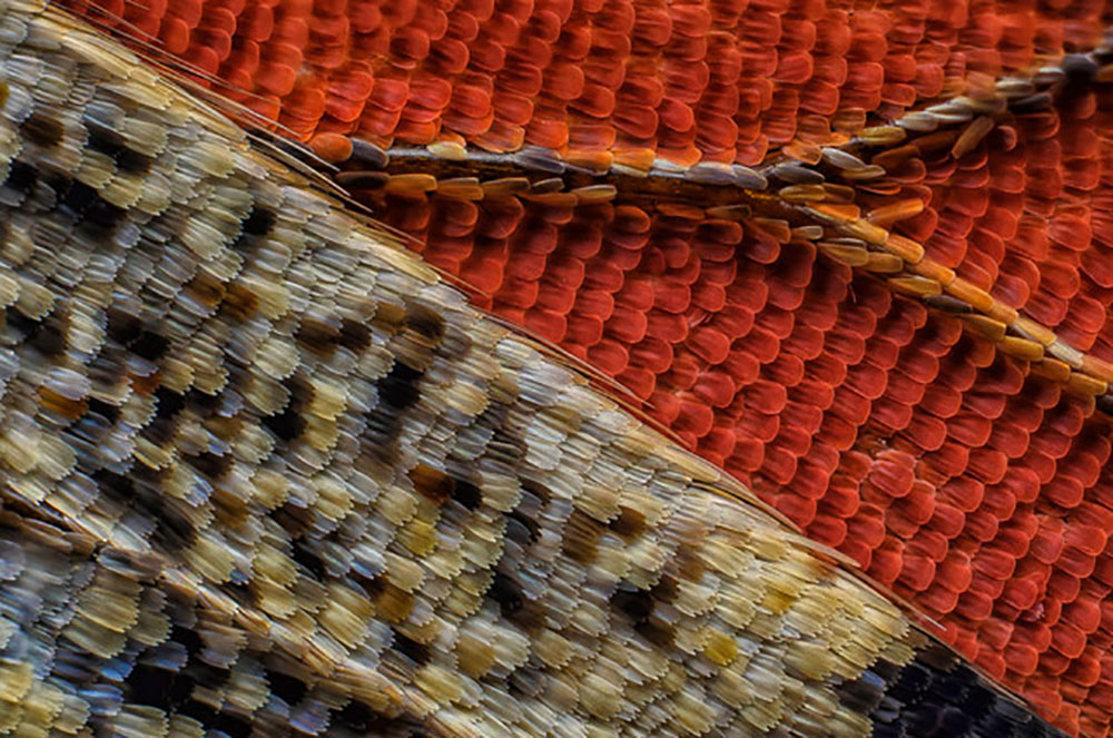
18th Place.
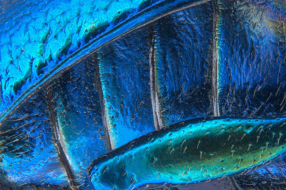
Pia Scanlon took Parts of wing-cover (elytron), abdominal segments and hind leg of a broad-shouldered leaf beetle (Oreina cacaliae).
19th Place.
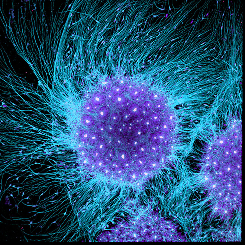
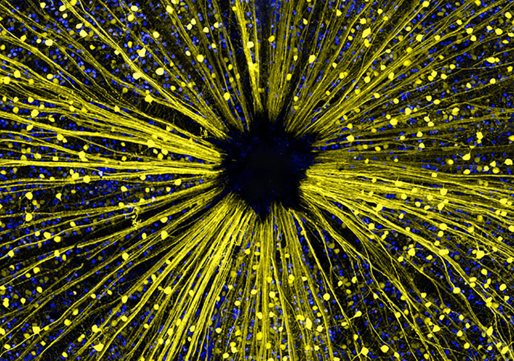
The techique was Confocal and the subject Human neural rosette primordial brain cells, differentiated from embryonic stem cells in the culture dish (used to study brain development and Huntington’s disease).
10th Place.
From Haverfordwest, Pembrokeshire, United Kingdom, this piece was taken by Michael Crutchley.
You can see the rest of the categories at www.nikonsmallworld.com


Loading…